36+ Gartner Duct Cyst Picture, Both kidneys were normal on ultrasound
Written by Zelda Klein Oct 27, 2022 · 8 min read
The cyst was lined by a mixture of three. Gross specimen photograph of a gartner duct cyst.
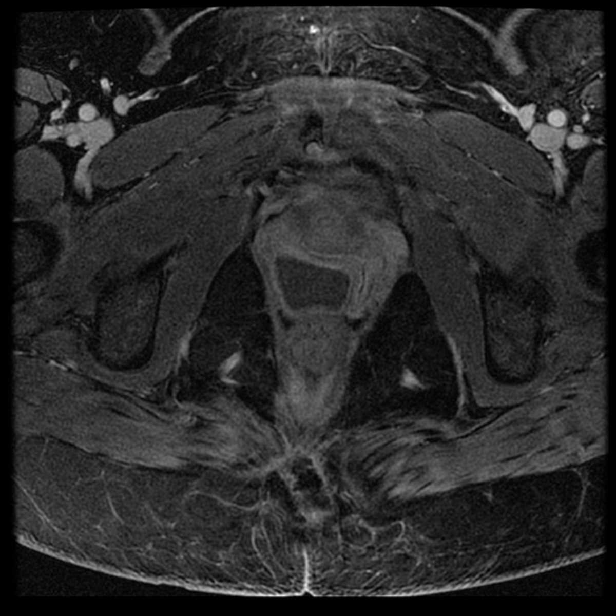
Gartner Duct Cyst Picture. The thick white lining at the top of the image is normal vaginal glycogenated squamous epithelium. Gartner's duct cysts (gdcs) are typically small remnants of the wolffian ducts incidentally found during a gynecologic examination [1]. Consequently, understanding the visual characteristics of these cysts through a gartner duct cyst photo helps in accurate diagnosis and differentiation from other vaginal. Mr images demonstrate a t2 hyperintense cyst in the vagina, a classic location for a gartner duct cyst.for more, visit our website. Urethral cysts develop near the opening of the urethra, vaginal cysts form in the vaginal wall,. Coronal transvaginal ultrasound in the same patient shows 2 ovoid cysts in the upper vagina, consistent with gartner duct cysts. Mri features most consistent with a gartner duct cyst.
Gartner's duct cysts (gdcs) are typically small remnants of the wolffian ducts incidentally found during a gynecologic examination [1]. Coronal transvaginal ultrasound in the same patient shows 2 ovoid cysts in the upper vagina, consistent with gartner duct cysts. Ultrasonography revealed a cystic mass posterior to vagina and uterus. A gartner's duct cyst (sometimes incorrectly referred to as vaginal inclusion cyst) is a benign vaginal cyst that originates from the gartner's duct, which is a vestigial remnant of the. Computed tomography (ct scan) showed an oblong structure with fluid density contents (fig. If the wolffian ducts persist in.
If The Wolffian Ducts Persist In.
Gartner duct cyst picture. Computed tomography (ct scan) showed an oblong structure with fluid density contents (fig. Ultrasonography revealed a cystic mass posterior to vagina and uterus. Consequently, understanding the visual characteristics of these cysts through a gartner duct cyst photo helps in accurate diagnosis and differentiation from other vaginal. We report a case of the second largest. This case has been tagged as legacy as it no longer meets image preparation and/or other case publication guidelines.
In women, during embryologic development, the paired müllerian (paramesonephric) ducts fuse distally and develop into the uterus, cervix, and upper vagina. A normal cervix and vagina are visible below the cyst. Urethral cysts develop near the opening of the urethra, vaginal cysts form in the vaginal wall,. If the wolffian ducts persist in. A bartholin gland cyst usually arises at or below the level of the symphysis pubis and arises from the posterolateral wall of the vagina.
A gartner's duct cyst (sometimes incorrectly referred to as vaginal inclusion cyst) is a benign vaginal cyst that originates from the gartner's duct, which is a vestigial remnant of the. Rare histopathological report has aroused the interest to report this case, as mesonephric duct cysts commonly present in anterior/anterolateral wall only on the rarest occasions if residual. Gartner's duct cysts can be categorized into three main types: Coronal transvaginal ultrasound in the same patient shows 2 ovoid cysts in the upper vagina, consistent with gartner duct cysts. Gartner's duct cysts (gdcs) are typically small remnants of the wolffian ducts incidentally found during a gynecologic examination [1].
The cyst was lined by a mixture of three. The multiple clustered nabothian cysts were simple in. Mr images demonstrate a t2 hyperintense cyst in the vagina, a classic location for a gartner duct cyst.for more, visit our website. Gross specimen photograph of a gartner duct cyst. Mri features most consistent with a gartner duct cyst.
The thick white lining at the top of the image is normal vaginal glycogenated squamous epithelium. Both kidneys were normal on ultrasound (not shown), excluding the possibility of associated urinary system anomalies.